

Leiden University Medical Center, the Netherlands
D. Jansma, A.C. Kraima, M.C. DeRuiter
Viewers partially based on the datasets of the Visible Human Project
-
Generation process of the UMCU pelvic datasetA proper specimen was selected to create a digital dataset of a female pelvis. The donated body belonged to a 69-year-old Dutch female, who had been suffering from multiple sclerosis for the last 20 years. There were no signs of external pelvic injuries and there was no history of pelvic dysfunctional complaints. The body was fixated in a 3% formaldehyde solution and preliminary MR images of the pelvis were obtained. The findings on MRI were examined by two experienced radiologists and confirmed to be free of anatomical abnormalities. The specimen was placed into a freezer at -25 C. The pelvis was separated and then cut into a block of 45 cm x 15 cm x 16 cm (lateral, ventro-dorsal and cranio-caudal, respectively), containing anatomically the lesser pelvis. The final block was constrained to 15 cm as this was the maximal size that could be sliced in the cryomicrotome (PMV 450; LKB Instruments, Stockholm, Sweden). Then, the pelvis was embedded in carboxymethylcellulose gel, frozen again at -25 C and transversally milled at 25 µm intervals. After wiping away frost, every third sectioned surface was photographed using a digital camera (Nixon D1X; Nikon Corporation; Chiypodaku, Tokyo, Japan) with a resolution of 3,040 x 1,961 pixels. After milling the complete pelvis, a series of 2051 images were acquired with a single pixel size of 82 µm. The images were saved as TIFF files and each image toke up 17.5 MB in size, resulting in all 35.9 GB. Coronal and sagittal images were reproduced from the transverse digital dataset using “Enhanced Multi Planar Reformatting Along Curves” software (E-MAC group, Department of Information and Computing Sciences, University of Utrecht, the Netherlands). In this way, a series of 1744 coronal images (2,648 x 2,048 pixels) and 2648 sagittal images (1,744 x 2,048 pixels) were acquired. Each image was saved as a BMP file (15.9 MB per coronal image and 10.5 MB per sagittal image), resulting in an additional dataset of 55.4 GB. After completing this entire process, structures of interest were able to be outlined using Amira 4.0 software.
Tables ( Review under submission: Towards a highly-detailed 3D pelvic model: approaching an ultra-specific level for surgical simulation and anatomical education. )

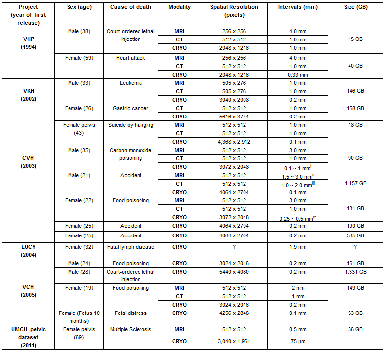
Table 1: Overview of features of the Visible Human Datasets (whole body and pelvis).
VHP: Visible Human Project, VKH: Visible Korean Human, LUCY: Stanford University model, CVH: Chinese Visible Human, VCH: Virtual Chinese Human. UMCU pelvic dataset: University Medical Center Utrecht pelvic dataset. i 0.5 mm intervals for the head and neck region, 0.1 mm intervals for the skull base and 1.0 mm intervals for other regions. ii 1.5 mm intervals for the head and neck region and 3.0 mm intervals for other regions. iii 1.0 mm intervals for the head and 1.0 mm intervals for other regions. iv 0.25 mm intervals for the head, 0.5 mm intervals for the trunk and 1.0 mm intervals for other regions.

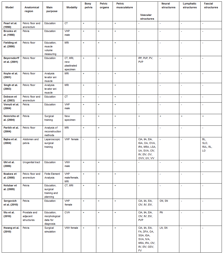
Table 2: Overview of all three-dimensional models concerning the pelvis or pelvic contents.
Abbreviations vascular structures. CIA: common iliac artery, IIA: internal iliac artery, EIA: external iliac artery, FA: femoral artery, DFA: deep femoral artery, OA: obturator artery, OVA: ovarian artery, SGA: superior gluteal artery, IGA: inferior gluteal artery, SVA: superior vesical artery, IVA: inferior vesical artery, MRA: middle rectal artery, IPA: internal pudendal artery, LSA: lateral sacral artery, UA: uterine artery, CIV: common iliac vein, IIV: internal iliac vein, EIV: external iliac vein, GSV: great saphenous vein, FV: femoral vein, OV: obturator vein, OVV: ovarian vein, UV: uterine vein, VV: vesical vein, IPP: internal pudendal venous plexus, PUP: periurethral venous plexus, PV: penile vein, PVP: prostatovesicular plexus. Abbreviations neural structures. ON: obturator nerve, LN: lumbar nerves, SN: sacral nerves, PN: prostatic nerves. Abbreviations fascial structures. BL: broad ligament, SLO: suspensory ligament of ovary, RUL: recto-uterine ligament, RL: round ligament, LO: ligament of ovary.